
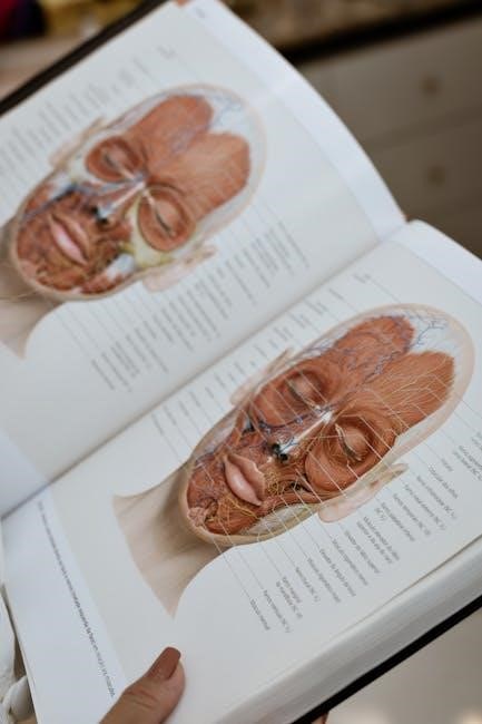
Human anatomy & physiology laboratory manuals are crucial resources‚ introducing students to core concepts and applied activities.
These manuals visualize structures‚ relating them to functions‚ and are designed for healthcare programs‚ offering extensive experiments.
Defining Anatomy
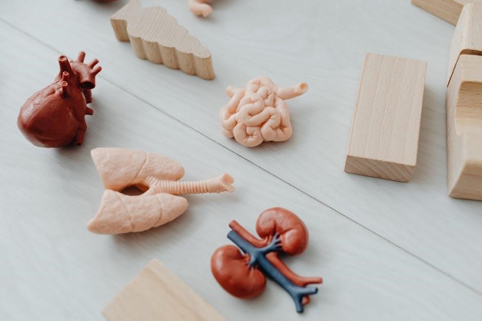
Anatomy‚ as explored within human anatomy & physiology laboratory manuals‚ fundamentally concerns itself with the structural organization of the human body. These manuals facilitate a detailed examination of both gross and microscopic structures‚ emphasizing a systematic understanding of bodily systems.
The study delves into identifying and describing the locations of various organs and tissues‚ utilizing dissections and microscopic observations.
Laboratory exercises within these manuals actively engage students in visualizing anatomical components‚ building a foundational knowledge base for comprehending physiological processes. The focus remains on precisely defining the body’s architecture.
Defining Physiology
Physiology‚ as presented in human anatomy & physiology laboratory manuals‚ investigates the functions of the human body and its constituent parts. These manuals move beyond structural descriptions to explore the dynamic processes occurring within living organisms.

Experiments and applied activities focus on understanding how organs and tissues operate‚ interact‚ and maintain homeostasis.
Students analyze mechanisms governing processes like respiration‚ circulation‚ and nerve impulse transmission. The laboratory component reinforces theoretical knowledge‚ bridging the gap between anatomical structure and functional performance‚ ultimately revealing how the body works.
The Relationship Between Structure and Function
Human anatomy & physiology laboratory manuals heavily emphasize the inseparable link between structure and function. These resources demonstrate how a body part’s form directly dictates its capabilities.
Laboratory exercises are designed to help students visualize anatomical structures and then relate those structures to specific physiological processes.
Understanding this connection is paramount; for example‚ the manual illustrates how the alveoli’s structure facilitates efficient gas exchange. This principle underscores the core tenet of the course – form truly follows function.
The Language of Anatomy
Laboratory manuals begin with “The Language of Anatomy‚” establishing foundational terminology for describing body structures and their relationships effectively.
Anatomical Position
Human anatomy & physiology laboratory manuals consistently emphasize the importance of the anatomical position as a standardized reference point. This universally accepted posture – standing erect‚ feet slightly apart‚ palms facing forward with thumbs out – allows for precise and consistent descriptions of body parts and their locations.
Understanding this position is fundamental because directional terms and regional designations are always referenced from this baseline. Manuals utilize diagrams and exercises to reinforce comprehension‚ ensuring students can accurately describe anatomical relationships regardless of the body’s actual orientation. This foundational knowledge is critical for effective communication within the field.
Directional Terms
Human anatomy & physiology laboratory manuals dedicate significant attention to directional terms‚ essential for accurately describing anatomical relationships. These terms – superior‚ inferior‚ anterior‚ posterior‚ medial‚ lateral‚ proximal‚ and distal – provide a common language for locating structures relative to one another.
Manuals often include exercises where students apply these terms to diagrams and models‚ solidifying their understanding. Mastering this terminology is crucial for interpreting anatomical descriptions‚ understanding clinical reports‚ and effectively communicating within healthcare settings. Consistent practice‚ as facilitated by lab manuals‚ is key to fluency.
Regional Terms
Human anatomy & physiology laboratory manuals systematically introduce regional terms‚ categorizing the body into specific areas like cephalic (head)‚ thoracic (chest)‚ abdominal (belly)‚ and appendicular (limbs). These terms offer a broader organizational framework than directional terms‚ allowing for focused study of interconnected structures within a defined region.
Lab exercises frequently involve identifying structures within these regions on anatomical models or cadavers. Understanding regional anatomy is vital for clinicians‚ enabling them to efficiently locate and assess specific body areas. Manuals reinforce this knowledge through detailed illustrations and practical applications.
Microscopy and Histology
Laboratory manuals dedicate sections to microscopy‚ detailing microscope types and slide preparation techniques for histological study of tissues.
Types of Microscopes
Human anatomy & physiology laboratory manuals thoroughly cover various microscope types essential for histological examination. These resources detail the principles behind light microscopy‚ the most commonly used technique for viewing cells and tissues.
Students learn about magnification‚ resolution‚ and proper illumination techniques. Manuals often introduce more advanced microscopy methods‚ potentially including phase contrast‚ interference‚ and even electron microscopy‚ though practical application may focus on light microscopy due to accessibility.
Understanding the strengths and limitations of each type is crucial for accurate observation and interpretation of biological structures;
Preparing Microscopic Slides
Human anatomy & physiology laboratory manuals dedicate significant attention to proper slide preparation techniques. Students learn essential skills like tissue sectioning‚ staining‚ and mounting to create specimens suitable for microscopic observation.
Detailed protocols guide the process of fixing‚ embedding‚ and sectioning tissues to achieve optimal thinness for light transmission. Manuals emphasize the importance of various staining methods‚ such as hematoxylin and eosin‚ to highlight cellular structures.
Proper slide preparation is paramount for accurate visualization and interpretation of histological features.
Basic Tissue Types
Human anatomy & physiology laboratory manuals systematically cover the four basic tissue types: epithelial‚ connective‚ muscle‚ and nervous. Students learn to identify these tissues under the microscope‚ recognizing their unique structural characteristics and functional roles.
Manuals provide detailed descriptions and illustrative images of various subtypes within each tissue category‚ such as different epithelial arrangements or connective tissue matrices.
Laboratory exercises often involve examining prepared slides and identifying tissue types in different organs‚ reinforcing the link between structure and function.
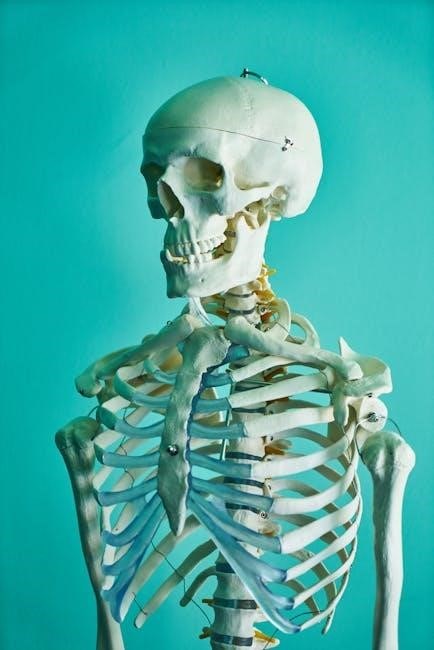
The Skeletal System
Laboratory manuals guide students through bone structure identification‚ classification‚ and articulation studies‚ enhancing understanding of the skeletal system’s form and function.
Bone Structure and Function
Human anatomy & physiology laboratory manuals dedicate significant sections to exploring bone structure‚ detailing both gross and microscopic anatomy. Students utilize these resources to identify key bone components – compact and spongy bone‚ periosteum‚ and marrow – through detailed illustrations and practical exercises.
These manuals emphasize the multifaceted functions of the skeletal system‚ extending beyond support and movement. They cover bone’s role in mineral storage‚ blood cell formation (hematopoiesis)‚ and protection of vital organs.
Laboratory activities often involve examining bone tissue under a microscope‚ analyzing bone composition‚ and understanding how structure directly relates to its physiological roles.
Bone Classification
Human anatomy & physiology laboratory manuals systematically categorize bones based on their shapes – long‚ short‚ flat‚ irregular‚ and sesamoid. These manuals provide detailed diagrams and examples‚ enabling students to accurately identify each type within the skeletal system.
Practical exercises often involve classifying specific bones‚ reinforcing understanding of the relationship between shape and function. The manuals explain how each bone class is uniquely suited to its role in the body‚ whether it’s providing leverage (long bones) or protection (flat bones).
Students learn to correlate bone classification with anatomical location and biomechanical properties.
Skeletal Articulations
Human anatomy & physiology laboratory manuals dedicate significant attention to skeletal articulations‚ detailing the diverse range of joint types – fibrous‚ cartilaginous‚ and synovial; These manuals utilize detailed illustrations and descriptions to clarify the structural characteristics of each joint category.
Laboratory exercises commonly involve identifying and classifying joints found in skeletal models or diagrams‚ emphasizing the correlation between structure and range of motion. Students learn to differentiate between various synovial joint subtypes‚ like hinge‚ pivot‚ and ball-and-socket joints.
The manuals explain how articulation impacts skeletal movement.

The Muscular System
Laboratory manuals for human anatomy & physiology explore muscle tissue types and contraction‚ featuring experiments to identify major muscle groups and their functions.
Muscle Tissue Types
Human anatomy & physiology laboratory manuals dedicate significant sections to exploring the diverse types of muscle tissue found within the human body. These resources typically guide students through microscopic observation and identification of skeletal‚ smooth‚ and cardiac muscle tissues.
Exercises often involve examining prepared slides to discern key characteristics like striations‚ cell shape‚ and the presence of multiple nuclei. The manuals emphasize the functional differences between these tissue types – skeletal muscle for voluntary movement‚ smooth muscle for involuntary actions within organs‚ and cardiac muscle exclusively for heart contractions.
Practical applications and experiments help students correlate microscopic structure with macroscopic function‚ solidifying their understanding of how each muscle type contributes to overall physiological processes.
Muscle Contraction
Human anatomy & physiology laboratory manuals thoroughly investigate the mechanisms behind muscle contraction‚ a fundamental physiological process. These manuals often include experiments demonstrating the sliding filament theory‚ utilizing models or simulations to illustrate actin and myosin interactions.
Students typically analyze the roles of calcium ions‚ ATP‚ and nerve impulses in initiating and sustaining contractions. Practical exercises might involve measuring muscle fatigue or observing the effects of different stimuli on muscle response.
The manuals bridge the gap between microscopic events at the sarcomere level and macroscopic observable muscle actions‚ reinforcing comprehension of neuromuscular function.
Major Muscle Groups
Human anatomy & physiology laboratory manuals dedicate significant attention to identifying and understanding major muscle groups throughout the body. These manuals commonly feature dissection exercises‚ requiring students to locate and palpate muscles on anatomical models or preserved specimens.
Students learn to categorize muscles based on their function – flexors‚ extensors‚ abductors‚ and adductors – and their location within specific body regions.
Practical activities often involve analyzing muscle origins‚ insertions‚ and actions‚ solidifying knowledge of how these groups contribute to movement and posture.
The Nervous System
Laboratory manuals guide students through neuron structure and function‚ brain and spinal cord anatomy‚ utilizing models and exercises for comprehension.
Neuron Structure and Function
Human anatomy & physiology laboratory manuals dedicate significant sections to exploring the intricate world of neurons. These resources typically guide students through identifying key neuronal components – dendrites‚ the cell body (soma)‚ the axon‚ and synaptic terminals – using microscopic images and diagrams.
Practical exercises often involve tracing the path of nerve impulses‚ demonstrating how signals are transmitted along the axon and across synapses. Manuals emphasize the functional roles of each structure‚ explaining how they contribute to communication within the nervous system. Students learn about action potentials‚ neurotransmitters‚ and the importance of myelin sheaths in accelerating signal transmission‚ solidifying their understanding through hands-on activities.
Brain Anatomy
Human anatomy & physiology laboratory manuals provide detailed explorations of brain anatomy‚ often utilizing models‚ diagrams‚ and preserved specimens. Students typically identify major brain regions – cerebrum‚ cerebellum‚ brainstem – and their subdivisions‚ like the frontal‚ parietal‚ temporal‚ and occipital lobes.
Labs frequently involve dissecting sheep brains to visualize internal structures‚ enhancing spatial understanding. Manuals emphasize the functional localization within the brain‚ correlating specific areas with sensory‚ motor‚ and cognitive functions. Exercises may include tracing pathways and identifying cranial nerves‚ solidifying knowledge through practical application and reinforcing the relationship between structure and function.
Spinal Cord Anatomy
Human anatomy & physiology laboratory manuals guide students through the intricate anatomy of the spinal cord‚ often utilizing models and cross-sectional diagrams. Labs focus on identifying key structures like the dorsal and ventral horns‚ gray and white matter‚ and the central canal.
Students learn to differentiate between spinal nerves and their roots‚ tracing pathways for sensory and motor information. Manuals frequently include exercises on identifying the components of a reflex arc‚ demonstrating the spinal cord’s role in rapid responses. Dissection‚ when available‚ provides a tactile understanding of the cord’s organization and protective meningeal layers.
The Cardiovascular System
Laboratory manuals aid in studying heart anatomy‚ blood vessels‚ and blood composition through dissections and microscopic observations of blood samples.
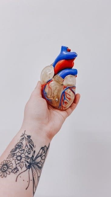
Heart Anatomy
Human anatomy & physiology laboratory manuals provide detailed exercises focused on dissecting and identifying the various components of the heart. Students learn to distinguish chambers – atria and ventricles – and valves‚ understanding their crucial roles in unidirectional blood flow.
These manuals often include activities to trace the pathway of blood through the heart‚ examining the coronary arteries and veins supplying cardiac muscle. Microscopic slides allow observation of cardiac muscle tissue‚ revealing its unique structure;
Furthermore‚ students analyze the heart’s conduction system‚ learning about the sinoatrial and atrioventricular nodes‚ and their impact on heart rate and rhythm.
Blood Vessels
Human anatomy & physiology laboratory manuals dedicate significant sections to blood vessel identification and function. Students dissect and examine arteries‚ veins‚ and capillaries‚ noting structural differences relating to pressure and oxygenation.
Labs often involve tracing major arteries and veins through anatomical models or preserved specimens‚ understanding their distribution and connections. Microscopic slides reveal the layered structure of vessel walls – tunica intima‚ media‚ and externa – and their composition.
Activities explore blood flow dynamics and the role of vessels in regulating blood pressure‚ crucial for cardiovascular health.
Blood Composition
Human anatomy & physiology laboratory manuals extensively cover blood composition‚ guiding students through identifying blood components under a microscope. Labs typically involve preparing blood smears and staining them to visualize erythrocytes (red blood cells)‚ leukocytes (white blood cells)‚ and thrombocytes (platelets).
Students learn to differentiate between various types of leukocytes – neutrophils‚ lymphocytes‚ monocytes‚ eosinophils‚ and basophils – based on their morphology and function. Hematocrit and hemoglobin measurements are common exercises‚ demonstrating quantitative aspects of blood composition.
Manuals emphasize the role of each component in maintaining homeostasis.

The Respiratory System
Laboratory manuals aid in understanding respiratory structures through dissection and modeling‚ exploring pulmonary ventilation and gas exchange mechanisms effectively.
Respiratory Structures
Human anatomy & physiology laboratory manuals provide detailed explorations of respiratory structures‚ crucial for understanding how we breathe. These manuals often incorporate dissection exercises focusing on the nasal cavity‚ pharynx‚ larynx‚ trachea‚ bronchi‚ and lungs.
Students utilize models and diagrams to identify each component and trace the pathway of air. Practical activities may involve examining lung tissue under a microscope‚ observing alveolar structure‚ and investigating the mechanics of breathing.
The manuals emphasize the relationship between structure and function‚ illustrating how each part contributes to efficient gas exchange‚ vital for sustaining life.
Pulmonary Ventilation
Human anatomy & physiology laboratory manuals dedicate significant attention to pulmonary ventilation‚ the process of moving air into and out of the lungs. These manuals frequently include experiments to demonstrate the mechanics of breathing‚ utilizing spirometers to measure lung volumes and capacities.
Students analyze data to assess respiratory efficiency and identify potential abnormalities. Activities often involve examining the roles of the diaphragm and intercostal muscles in altering thoracic volume.
The manuals emphasize the principles of pressure gradients and airflow‚ illustrating how ventilation ensures adequate oxygen intake and carbon dioxide removal.
Gas Exchange
Human anatomy & physiology laboratory manuals thoroughly explore gas exchange‚ the vital process occurring in the lungs and tissues. These manuals often incorporate simulations or experiments to demonstrate the diffusion of oxygen and carbon dioxide across the alveolar and capillary membranes.
Students investigate factors influencing gas exchange efficiency‚ such as partial pressure gradients and membrane permeability.
Activities may involve analyzing the impact of various conditions‚ like anemia or emphysema‚ on oxygen transport. The manuals reinforce understanding of how structure dictates function in this critical physiological process.